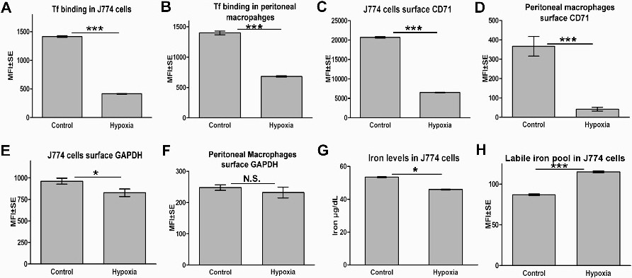
Fig. 5. Macrophages are not programmed for enhanced iron acquisition in response to hypoxia. (A&B) In contrast to the results in K562 cells, transferrin binding is significantly decreased in macrophages subjected to hypoxia: 24 hrs hypoxia treated J774 (A) and mouse peritoneal macrophage (B) cells were assayed for transferrin binding by flow cytometry. For both cell types p<0.0001, n=104. (C&D) Cell surface CD71 expression in macrophages is decreased upon exposure to hypoxia: J774 (C) and mouse peritoneal macrophages in cell culture (D) exposed to 24 hrs hypoxia were stained for surface expression of TfR1 (CD71) and evaluated by flow cytometry. Results were compared to normoxic cells. Data is presented as mean fluorescence intensity ± SE. p<0.0001, n=104. (E&F) Hypoxia exposed macrophages do not deploy GAPDH on their surface: J774 (E) and mouse peritoneal macrophage (F) cells in culture were subjected to 24 hrs of hypoxia and then evaluated for surface expression of GAPDH. No significant increase in deployment of GAPDH on plasma membrane is observed. Data is presented as mean fluorescence intensity ± SE, p<0.05 for J774 and p>0.05 for peritoneal macrophages, n=104. (G) Decreased cellular iron levels in hypoxic J774 cells as hypoxia induces macrophage cells to release iron, p<0.05, n=3. (H) Calcein quenching assay reveals a depleted labile iron pool. As presence of iron quenches calcein florescence the observed increase in signal from hypoxic J774 cells indicates a depleted labile iron pool. Data is presented as mean fluorescence intensity ± SE, p <0.0001, n=104.